A 72-year-old male with right upper lung primary non-small cell lung cancer was referred to us as an inpatient in one of our partner hospitals. He was status post right upper lobe lobectomy with thoracic lymph node resection by the thoracic surgeon. Post-operative hospital course was complicated by a persistent right chylous pleural effusion despite initiating octreotide therapy and a medium-chain triglyceride oil diet of 80 cc/day for over 10 days. Our interventional radiology APP, Anais Pedoussaut, PA-C, was consulted for evaluation. The patient subsequently underwent a lymphangiogram with thoracic duct embolization with coils and glue with our interventional radiologists, Dr. Kumar Shah and Dr. Adam Checkver, along with our ESIR resident, Dr. Geoffrey Lindblad.
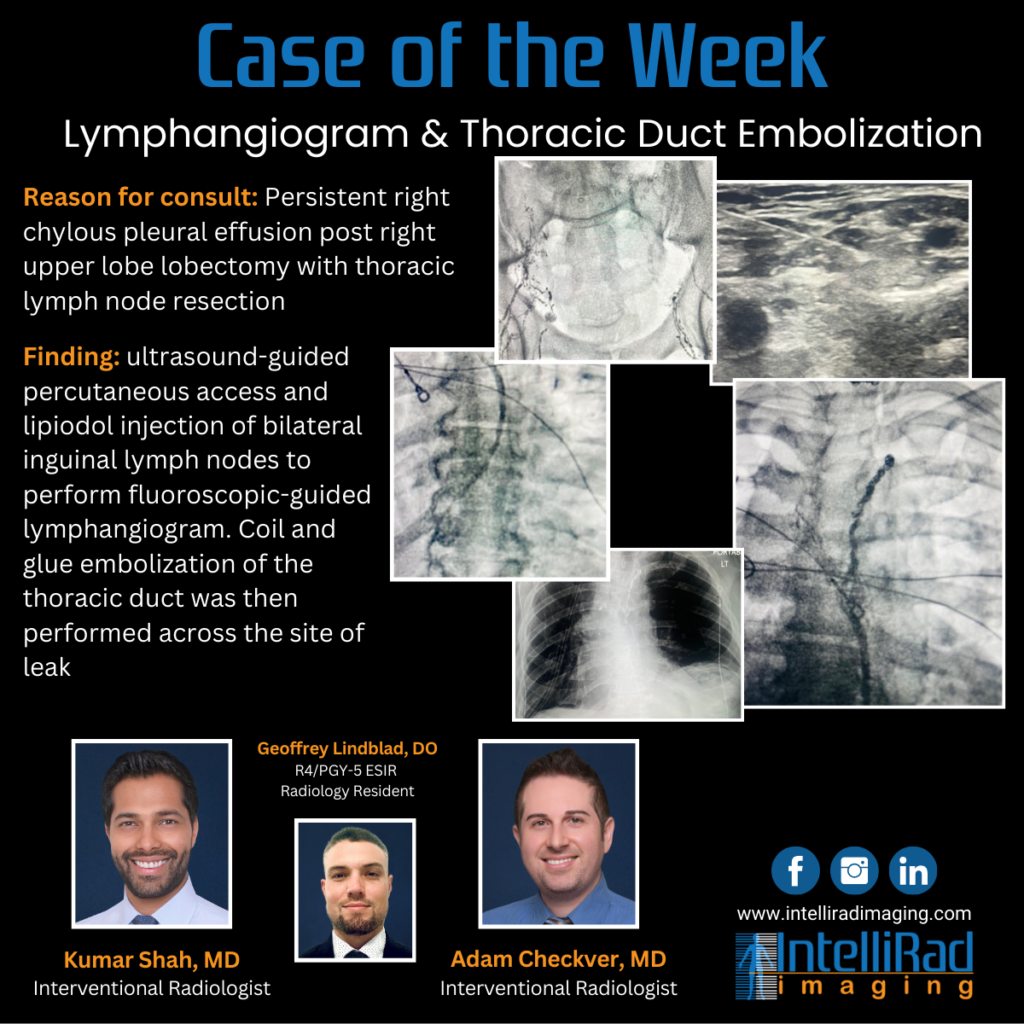
The procedural details includes ultrasound-guided percutaneous access and lipiodol injection of bilateral inguinal lymph nodes to perform fluoroscopic-guided lymphangiogram. Once the lymphatic system was visualized and mapped out, the cisterna chyli was directly punctured under fluoroscopic guidance. DSA imaging demonstrated the thoracic lymphatic duct in the chest with an area of discontinuity/leak in the right mid to upper chest. A coil and glue embolization of the thoracic duct was then performed across the site of leak.
Right pleural chest tube output decreased immediately following the embolization with resolution of the chylous effusion. Patient was discharged a few days later.